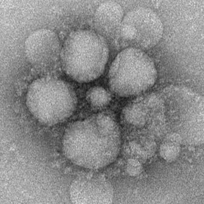
Coronavirus Actual Image Under Microscope. This isn't quite as sharp as the first one, but you can see the spikes on the surface Viruses in the coronavirus family only have small differences in their genome, with only five nucleotide differences between three of the viruses. How does this tiny particle, that has turned the world upside down, actually work and what.
The images of the coronavirus have been taken by a team of ICMR-NIV scientists in Pune.
They were made with scanning and transmission electron microscopes.
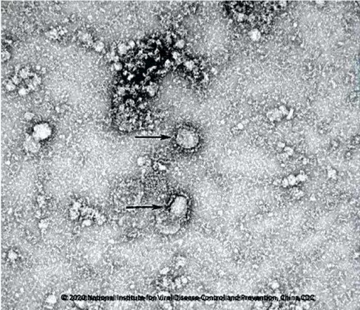
First image of new coronavirus released - CGTN
Why does the coronavirus spread so easily between people?

COVID-19: Treatment and Vaccine Development Move Ahead ...

3d model of coronavirus stock illustration. Illustration ...

Australia may be first country to find coronavirus cure ...

Corona Virus On Microscope - Never lost your place

Fonds d'écran Coronavirus au microscope électronique ...

health coronavirus virus covid-19 2019-nCoV under ...

Coronavirus Covid -19 Danger Cell Under The Microscope And ...
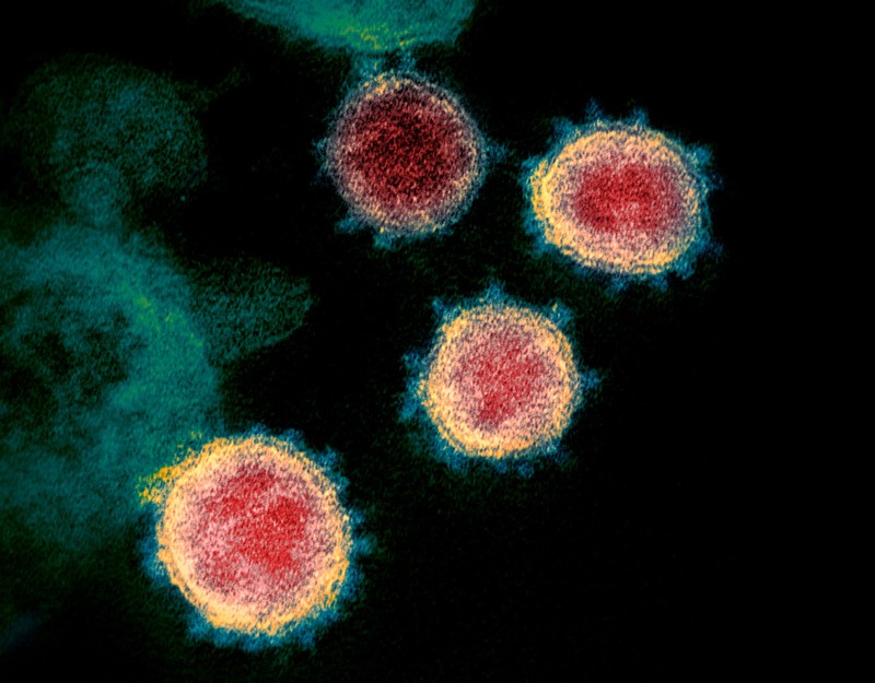
ResearchGate, the professional network for scientists. Jump to navigation Jump to search. This scanning electron microscope image shows the new coronavirus (yellow) among human cells (blue, pink and purple).