Covid Brain Image. Thousands of new, high-quality pictures added every day. Tumour patient says second operation might have been avoided had MRI image been taken.
The CT is normal or there are findings that indicate a non-infectious disease like congestive.
COVID working group of the Dutch Radiological Society.
Radiology, MRI
COVID-19 y coagulopatía, ¿qué sabemos hoy?

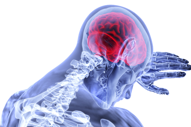
Scientists warn of potential wave of COVID-linked brain ...
Does CSF Antibody Testing Confirm Coronavirus in the Brain?
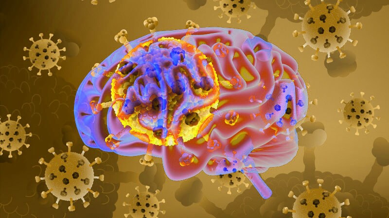
COVID-19 may harm both the brain and the lungs | PhillyVoice

Brain problems linked to even mild coronavirus infections ...

Fact check: COVID-19 nasal test doesn't swab blood-brain ...

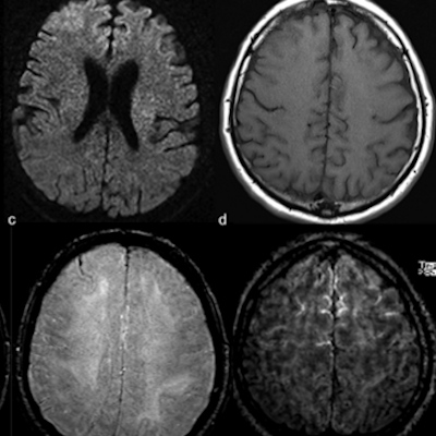
COVID-19 Neurological Manifestations and Brain Volumetrics

From Brain to Toes, The Covid-19 Symptoms you didn't know ...
Brain scans from Magnetic Resonance Imaging experiments (MRI) have been a popular choice with I am starting with image analysis and this is helpful. Brain Imaging: An Introduction presents diverse manifestations of brain disease as shown by neuroradiology. Researchers at the University of Oregon took advantage of an advanced brain imaging technique known as functional magnetic resonance.