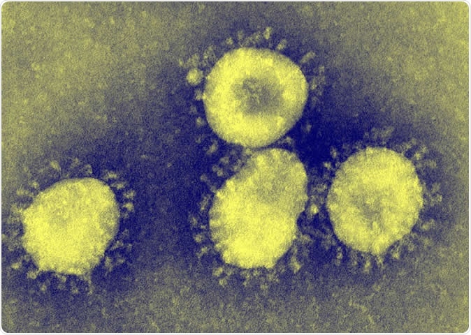
Image Coronavirus Under Microscope. For the most up-to-date news and information about the coronavirus pandemic, visit the WHO website. This isn't quite as sharp as the first one, but you can see the spikes on the surface Viruses in the coronavirus family only have small differences in their genome, with only five nucleotide differences between three of the viruses.

The image above was captured with a transmission electron microscope.
The horseshoe bat and the pangolin have both been implicated, but the precise sequence of events is unknown.
Anti-malaria drug being tested for efficacy against COVID-19
Here’s how nanoparticles could help us get closer to a ...

Image Of Flu COVID-19 Virus Cell Under The Microscope On ...

Pin en High Resolution Photo

Coronavirus explainer: Under the microscope | ABC News ...
First exclusive images of the virus under a microscope ...

Electron microscopy captures snapshot of structure ...

Coronavirus | New Scientist
Coronavirus covid-19 under the microscope. 3d illustration ...
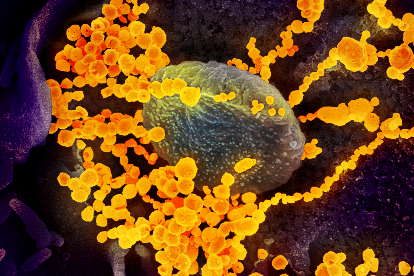
This scanning electron microscope image shows the new coronavirus (yellow) among human cells (blue, pink and purple). English: Coronaviruses are a group of viruses that have a halo, or crown-like (corona) appearance when viewed under an electron microscope. Additional specimens are being tested to learn more about this.